Cardiovascular disease is still the world’s biggest killer, and a major culprit is atherosclerosis—plaque buildup that narrows arteries and can trigger strokes. The carotid arteries (in the neck) are especially important: their shape and blood-flow patterns are strongly linked to where plaque forms and how it progresses. Clinicians would love to measure patient-specific blood flow in detail, because flow-related markers (like how strongly blood “scrubs” the vessel wall, or how much flow direction changes) are valuable risk indicators.
There’s a problem: the best non-invasive method for capturing full 3D, time-resolved blood-flow patterns—4D flow MRI—is expensive, time-consuming, and not widely available. It also suffers from measurement noise and can be tricky to acquire correctly. The alternative is computational fluid dynamics (CFD) simulations, which can produce physics-consistent flow estimates, but often require heavy computation, long runtimes, and careful choices about modeling details (mesh quality, boundary conditions, solver settings). Worse, different teams can get noticeably different CFD results for the same case, making comparisons hard.
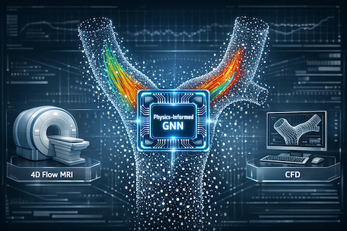
This paper proposes a third path: a fast surrogate model that predicts detailed blood-flow velocity fields directly from a patient’s artery geometry—using machine learning, but with a key twist: it’s physics-informed and symmetry-aware.
The core idea: learn blood flow from shape, but don’t let the model “cheat”
Blood flow in arteries depends heavily on:
-
the 3D geometry (how the vessel curves, narrows, branches)
-
the inflow conditions (how much blood enters, how variable it is)
The authors train a neural network to take a point-cloud representation of the carotid artery lumen (instead of a mesh) plus simple boundary-condition features, and output a 3D velocity vector at each point—essentially reconstructing the flow field.
Why point clouds? They avoid locking the model into a specific mesh connectivity and help reduce overfitting to how a surface was triangulated. It’s also a natural fit for models inspired by PointNet++ (a well-known architecture for learning on unordered 3D points).
Making it “physics-aware” in two ways
-
Symmetry (equivariance) built into the network
In real life, if you rotate or translate the artery in space, the flow pattern should rotate/translate the same way—no more, no less. The authors enforce this by using steerable, equivariant layers so the model respects these geometric symmetries by design. This typically improves data efficiency: you don’t need to see the same anatomy in every orientation to learn the right behavior.
-
Physics-informed training regularization
Even good MRI flow measurements can be noisy—especially near the vessel wall. To stop the model from learning noise as if it were “real” flow, the authors add training penalties that push predictions toward physically consistent behavior (based on the governing fluid dynamics). Importantly, they do this without requiring a heavy volumetric mesh.
They introduce a mesh-free discretization approach using local neighborhood queries (nearby points) to compute physics residuals efficiently. That’s a practical win: building high-quality volumetric meshes is hard and can explode compute/memory costs.
What they trained on (and why it matters)
A major departure from many prior studies: instead of training on synthetic CFD labels, they train primarily on in-vivo 4D flow MRI measurements from a moderately sized real dataset:
-
234 subjects
-
carotid bifurcation region
-
they focus on peak systole (the moment with the highest average velocity)
This is crucial because CFD-based “ground truth” is only as good as the modeling assumptions. By learning from real measurements, the model aims to reflect what’s observed in humans—not what a particular CFD pipeline produces.
Results: accurate, cleaner flow fields—and transferable knowledge
Across experiments, the model:
-
predicts flow fields that match measured 4D flow MRI well in direction and overall structure
-
often produces smoother, more plausible flow than noisy MRI in difficult cases (suggesting it’s learning the anatomy→flow relationship and treating noise as noise)
-
shows that adding equivariance improves adherence to physical constraints
-
shows that adding physics-informed regularization further reduces unphysical behavior (but if weighted too heavily, the model can collapse toward overly “average” predictions—so balance matters)
The most clinically interesting part: generalizing to a different imaging modality
The authors also test whether what the model learns from 4D flow MRI can transfer to black-blood MRI—a faster, widely used anatomical scan that doesn’t measure velocity.
They take vessel geometries derived from black-blood MRI and feed boundary-condition inputs (meant to mimic what could be measured with something like Doppler ultrasound). The model still produces flow estimates that qualitatively align with the true 4D flow MRI patterns for the same patients.
This suggests a realistic future workflow:
-
use widely available anatomical imaging (or potentially CT / ultrasound-based geometry)
-
get simple inflow info non-invasively
-
generate fast, patient-specific flow estimates
-
reserve expensive 4D flow MRI for complex or suspicious cases
Bottom line
This work argues that physics-informed, symmetry-aware graph/point-cloud neural networks can learn meaningful blood-flow prediction from real clinical measurements, reduce reliance on slow CFD, and extend hemodynamics estimation to settings where full flow MRI isn’t feasible. It’s a strong step toward making advanced flow-based cardiovascular risk assessment more scalable in real hospitals.
source: https://arxiv.org/pdf/2408.07110